MIWATCH tracking indium at the Australian Synchrotron

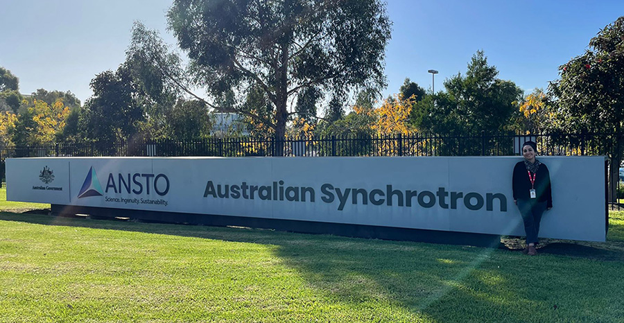
This month, Thomas Ray Jones from the School of Earth and Environmental Sciences (SEES) and I had a challenging, exciting and intensive two days at the Australian Synchrotron in Melbourne, Australia. Here, X-ray fluorescence microscopy (or XFM) beamline experiments were carried out as a part of my PhD which aims to identify potential hosts of the critical metal indium in mine waste to build a better understanding of its surficial cycling and geochemical mobility.
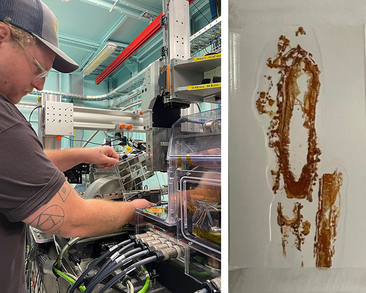
The XFM beamline experiments involved analysing a variety of indium-rich samples, including drill core, waste rock, sediments and precipitates, containing bulk indium ranging from 1 to 802 ppm. These samples were collected from two abandoned mines where I also found dissolved indium in open pit and sumps waters.
After running different XFM experiments (changing the spot size, energy and velocity), we collected incredible images at a resolution of 2 microns. These revealed complex textures of intergrown massive sulphide minerals, and the breakdown of sulphides to secondary minerals with notable entrapment of microorganisms with both base- and critical-metals identified.
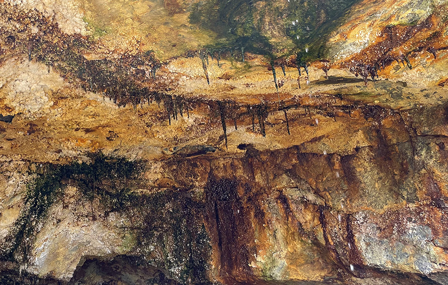
Right image: A thin section of an indium-rich dripstone.
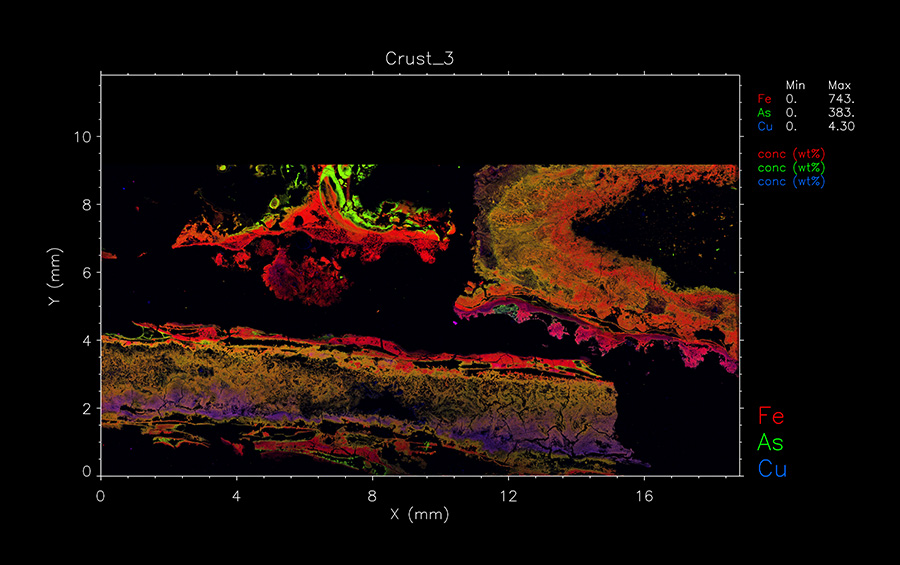
One challenge we faced- despite the high bulk indium contents in several samples, was that the indium signal being studied overlaps with those for other elements including tin, potassium, and calcium, making it challenging to obtain a clean signal of indium. However, based on previous scanning electron microscopy and mineral chemistry work (by laser ablation ICP-MS), we were able to confirm that the indium signal in the mineralised sample is overlapping with the indium-bearing mineral stannite.
In contrast, the indium signal in the dripstone sample overlapped with calcium, sulphur, and potassium, as well as the presence of microorganisms, indicating that secondary minerals may be trapping indium on their surfaces involving a biogeochemical process. The findings suggest that by undertaking a multiscale characterization approach we will get a better understanding of the trace distribution of indium.
This opens up new opportunities to trial emerging technologies where the indium overlap spectra/signal effect is much smaller (e.g. LIBS for which I has extensively tested field-portable devices).
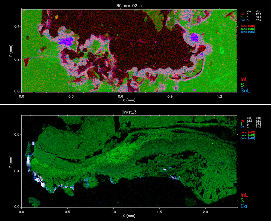
Top image: Mineralised sample (802 ppm In). Bottom image: Dripstone sample (175 ppm In).
These images are still under data analysis/review.
My plan is to process, in-depth, the new data collected from the XFM synchrotron beamline and complement them with data from other techniques to confirm indium cycling in mine waste environments. I can’t wait to share the next discoveries!
This research project was funded by synchrotron grant award round 2023/1, and it is kindly supported by the Geological Survey of Queensland (GSQ) under the New Economy Minerals Initiative (NEMI) program.