XANES mapping of cobalt speciation and mobility at the Australian Synchrotron
Beamtime at the Australian Synchrotron is highly competitive and this XANES experiment was the 3rd successfully awarded beamtime for the MIWATCH team in the last 14 months! Previously, the XFM (X-Ray Fluorescence Microscopy) beamline has been utilised by the MIWATCH team to spatially map cobalt and indium mobility in mine tailings. In this blog, PhD candidate Loren Nicholls shares how XANES technology can be applied to understanding cobalt speciation and secondary mineralogy in historical copper tailings.
XANES (X-ray absorption near-edge structure) mapping utilises the XFM beamline to detect the oxidation state of elements in a sample. When a sample is hit by the x-ray beam at an energy above its absorption edge, a core electron is excited and leaves a hole in the atomic core level which is then filled by an outer electron, releasing a ‘fluorescent’ x-ray. The absorption energy of an element changes with its oxidation state and the other atoms that surround it. XANES is a technique that changes the energy of the x-ray beam across this edge, in the case of cobalt from 7580 to 7850 eV, to show absorption features like pre-edge features, edge location, the white line zone and post edge features which are indicative of oxidation state and what other atoms it is bonded to. Figure 2 illustrates these features on a XANES spectra.
The reason for applying for XANES XFM beamtime was the complex nature of the copper tailings and the challenges this poses for characterisation. This material had both primary and secondary mineralogy, as well as compounds from mineral processing fluids, all of which have undergone oxidation via weathering and cyclical evaporation/saturation. The material is also incredibly fine grained, making it difficult to target individual grains. Finally, the material is pyrite rich, meaning a high Fe signature. The Fe spectral signature overlaps with Co and causes significant interference. By isolating the Co Kα edge from the Fe signature using XANES, we were able to definitively identify cobalt hosts.
The experiment was pretty novel for the XFM beamline, but the results turned out amazingly! We all geeked out on the beautiful images and cool signatures identified. Figure 4 shows two different species of cobalt; Co in oxide and Co in cobaltite.
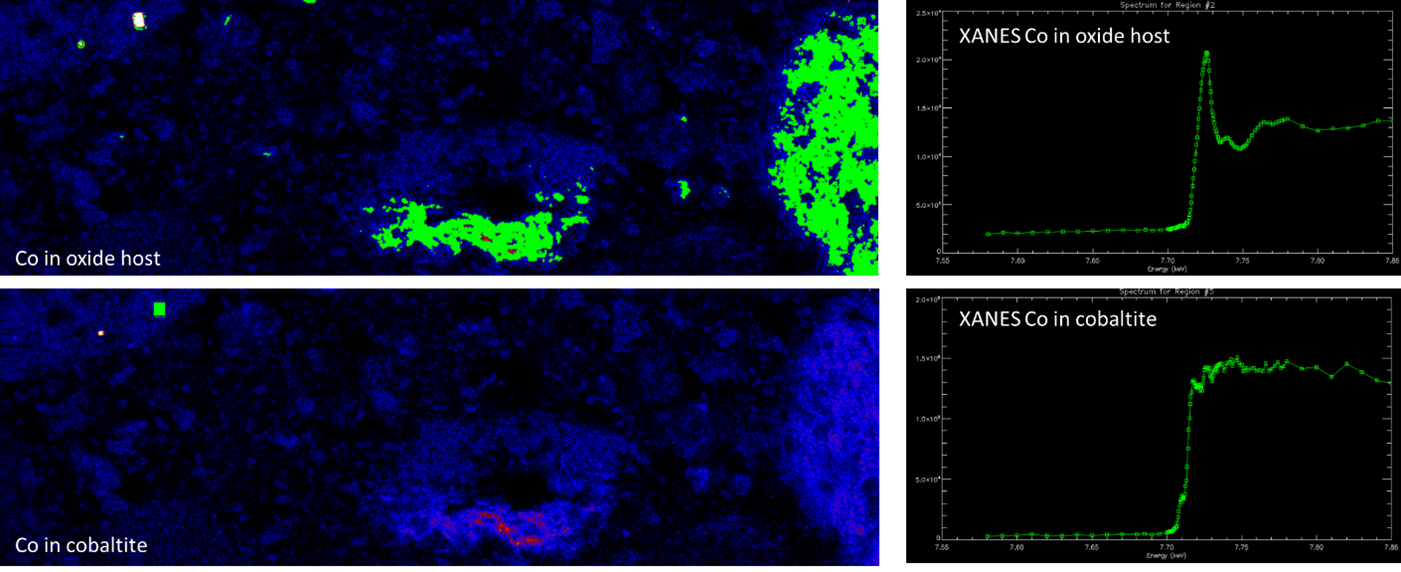
Right: XANES response of the green highlighted areas. The two different responses indicate different oxidation states and hosts of cobalt.
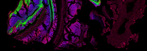
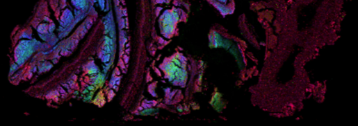
The XFM maps were also spectacular, showing the relationship between Co, Fe, Mn, Ca, Cu and As. Figures 5 and 6 show an RGB images of Fe-Mn-Co and Fe-Cu-Co for the same sample. Cobalt and copper predominantly occur in a secondary sulphate phase with copper in the centre and cobalt on the outer rim. Around the fractures there is an additional Mn-Co phase present.
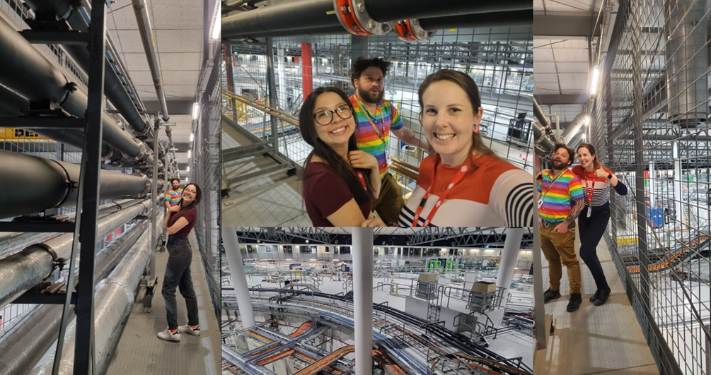
This was Loren’s first time at the synchrotron, Mayra’s second time and Tom’s umpteenth time, yet the facility still blows us all away. Everything runs like clockwork, there are procedures and protocols, very clear communication and well appointed guest rooms so that you can stay doing science all night! Andrew (beamline scientist) gave us a great after-hours tour of the facility and talked through his immense knowledge on all the different beamlines and their applications.